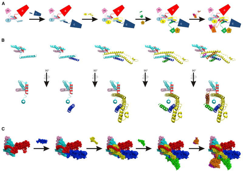
Figure 5. Main Assembly Pathway for Human-like eIF3.
(A) Cartoon scheme showing the ordered assembly of eIF3. Helical bundle formation is guided by the C-terminal helices of the indicated subunits. The (b,g,i) subcomplex assembles with eIF3a, independently of assembly of eIF3f and m with eIF3a, and is not depicted. Assembly of eIF3d with eIF3e is indicated, but is not dependent on the C-terminal helices of eIF3e.
(B) Two views of eIF3 helical bundle assembly. Subunits are added as in (A), except eIF3d is not included.
(C) Main assembly pathway of the human eIF3 octamer according to helical bundle assembly in (B), showing the available structural models for the subunits. Images in (B) and (C) are from the cryo-EM structure of human eIF3 (PDB: 5A5T) (des Georges et al., 2015). Subunits in all panels are colored as follows: eIF3a (red), eIF3c (blue), eIF3d (brown), eIF3e (green), eIF3f (cyan), eIF3h (yellow), eIF3k (magenta), eIF3l (orange), and eIF3m (light pink). See also Figures S2–S4.

