Figure 5.
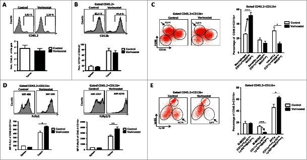
Vorinostat treatment increases the presence of macrophages while reducing M-MDSC in the TME of NBL tumors. Mice bearing 9464D tumors (6 mice/group) were treated with Vorinostat (150 mg/kg) for 3 consecutive days after which tumors were excised and single-cell suspensions were made. (A) Vorinostat does not alter total leukocyte infiltration of NBL tumors. The total tumor cell suspension was analyzed for the presence of CD45.2+ leukocytes. Representative data from three independent experiments. (B) Vorinostat does not alter myeloid cell presence in spleens and tumors. CD45.2+ leukocytes were gated and analyzed for the expression of CD11b. Representative data from three independent experiments are shown. (C) Vorinostat increases the presence of macrophages within the tumor infiltrating myeloid cells. CD45.2+CD11b+ tumor-infiltrating myeloid cells were gated and analyzed for the expression of CD11c, F4/80 and MHCII. Percentages of CD11cdimF4/80highMHCIIint macrophages, CD11chighF4/80dimMHCIIhigh DC and CD11clowF4/80lowMHCIIlow non-APC are depicted (*p < 0.05, ***p < 0.001). Representative data from three independent experiments are presented. (D) Vorinostat upregulates the expression of FcRγ1 and FcRγ2/3 on the cell surface of tumor-infiltrating myeloid cells. CD45.2+CD11b+ myeloid cells were gated and analyzed for the expression of FcRγ1 and FcRγ2/3 (*p< 0.05, **p< 0.01). Representative data from three independent experiments. (E) Vorinostat reduces M-MDSC within the tumor-infiltrating myeloid cells. CD45.2+CD11b+ myeloid cells were gated and analyzed for the expression of CD11c, Ly-6C, Ly-6G and MHCII. Percentages of and CD11cnegLy-6ChighLy-6GnegMHCIIlow M-MDSC, CD11clow/intLy-6CdimLy-6GhighMHCIIlow PMN-MDSC and CD11cint/highLy-6CnegLy-6GnegMHCIIint/high APCs are depicted (*p < 0.05; ***p < 0.001). Representative data from three independent experiments are shown.

