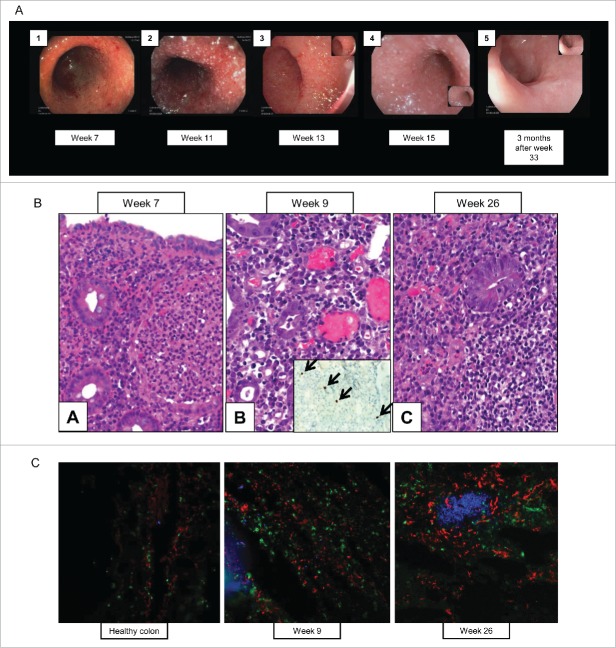
Figure 2.
(A) High-definition endoscopy. Pictures 1–4 of weeks 7, 11, 13, and 15 describe both in high definition white-light (3, 4) and in virtual chromoendoscopy (1, 2) an erythematous, granulomatous and oedematous mucosal inflammation of the rectosigmoid colon with a high mucosal vulnerability (spontaneous diffuse mucosal bleeding). Images 2 and 4 show a particular grainy white pattern suggesting an overlapped infectious origin confirmed by immunohistochemistry and microbiology as a CMV colitis which superposed and complicated the initial autoimmune T cell driven ipilimumab colitis. As a comparison, picture 5 taken 3 mo after week 33 shows an almost complete mucosal restitution with reminiscent erythema, hypervascularity and healed scars in the colon. (B) Immunohistochemistry of colon biopsies with CMV staining. [A]: pre-CMV initial biopsy showed prominent plasma cell-rich inflammatory infiltrates in the lamina propria and notable crypt abscess formation. [B]: CMV-stage: the crypt epithelium showed regenerative changes with large intranuclear inclusions, the lamina propria contained mixed infiltrates with prominent dilated capillaries. Inset: CMV was strongly positive in four nuclei in this field (arrows). [C]: post-CMV biopsy showed still prominent mononuclear cell infiltration but no neutrophilic crypt abscesses, note follicular hyperplasia. (C) Multi-epitope ligand cartography (MELC) of cryo-conserved colon tissue. Samples taken at two different time points and a control sample from healthy colon have been stained for different antibodies using the MELC-technique.19 Natural killer cells (CD56+) and CTLA-4 positive cells increased over time, and B cells (CD20+), CD8+ T-cells, memory T cells (CD45RO+) and CD45+ T cells additionally formed follicles. Depicted is an overlay (20X magnification) of NK cells (red), CD8+ T cells (green) and B cells (blue).

