Figure 1.
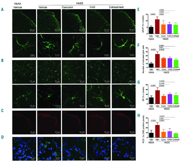
Glial activation, neuroinflammation and oxidative stress in spinal cords of sickle mice is ameliorated with curcumin and/or CoQ10. Analysis of activation in glial cells in L4-L5 superficial dorsal horn segments of female sickle mice after daily treatment with curcumin (HbSS Curc), CoQ10 (HbSS CoQ), or both (HbSS Cotreatment) for 4 weeks. Sickle mice (HbSS Vehicle) show increased levels of activated cells as compared to control mice (HbAA Vehicle) which is reduced by all treatments. Representative images of (A) astrocytes (GFAP, upper panel; lower panel showing a magnified field of activated astroglial cells), (B) activated microglia (Iba1 positive cells, upper panel; lower panel shows magnified fields of activated microglial cells, (C) neuropeptide SP, and (D) ROS (green) with DAPI counterstained nuclei (blue). Quantitative analysis of (E) GFAP immunoreactivity, (F) numbers of resting and activated microglial cells, (G) SP immunoreactivity, and (H) ROS levels expressed as percentage of DHE positive pixels. Each image is representative of images from 8 mice per treatment. Scale bars as indicated. Quantitation of fluorescence expressed as the mean value of the pixel density ± SEM of 3 nonadjacent fields per mouse, except (F) activated microglial expressed as number of immunoreactive cells per field. Significance was determined by one-way ANOVA with Bonferroni’s multiple comparison.

