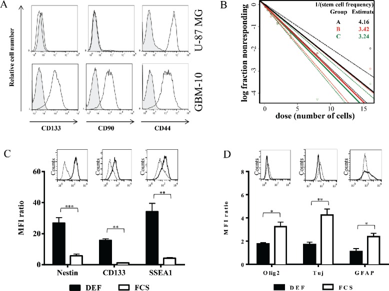
Figure 3.

Primary GBM-10 cells as cellular tools for the establishment of a physiological orthotopic human GBM graft model in NSG mice. (A) U-87MG cells and GBM-10 cells were analyzed by flow cytometry for CD133, CD90 and CD44 expression. Gray histograms correspond to isotype control mAbs. (B) GBM-10 cells were seeded with an initial concentration of 2 × 10³ cells/mL in 96-well plate. 15 d later the fraction of wells not containing neurospheres for each cell-plating density was calculated. (C–D) GBM-10 primary cells were cultured in DEF or in medium containing FCS and were analyzed by flow cytometry for the Nestin, CD133, SSEA1 (C), Olig2, Tuj and GFAP expression (D). Results are expressed as the median fluorescence intensity (MFI) ratio (MFI test/isotype control) (mean ± SEM, n = 3; *p <0.05, **p <0.005, ***p <0.0005). Inserts show representative histograms from one experiment of three performed.
