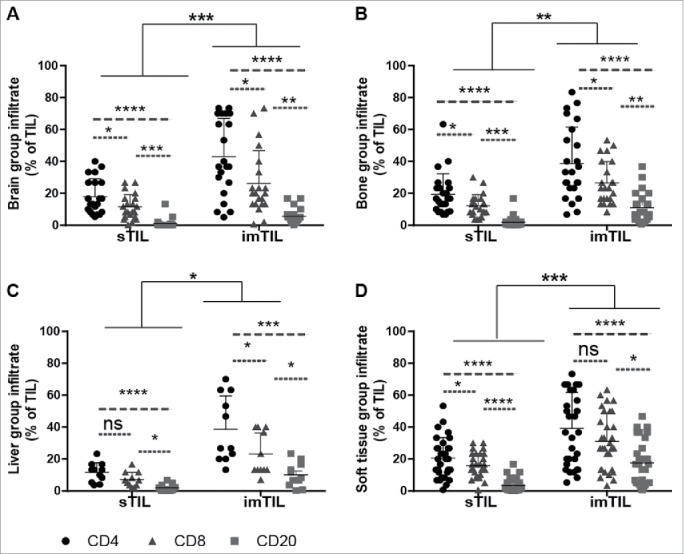
Figure 2.

Similar distribution of TIL with regard to the investigated primary tumor compartments and the lymphocyte subtypes but irrespectively of the site of distant metastasis. Scatter graphs show that within the primary tumor compartments (iTIL (not shown), sTIL and imTIL) invasive margin TIL (imTIL) are significantly increased when compared to stromal (sTIL) TIL irrespectively of the particular site (A–D) to which metastasis had occurred. For all three compartments (iTIL not shown) CD4+, CD8+ and CD20+ lymphocyte subsets always followed the same gradient with CD4+ lymphocytes being the most prominent, followed by CD8+ lymphocytes and at last CD20+ lymphocytes again irrespectively of the anatomical site of distant metastasis (A–D). Significances are displayed as follows: ns = p > 0.05, *p ≤ 0.05, **p ≤ 0.01, ***p ≤ 0.001 and ****p ≤ 0.0001.
