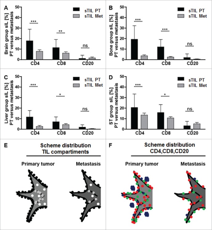
Figure 3.

Significantly more sTIL within the primary tumor than mTIL within the site of the corresponding metastasis. Bar graphs for CD4+, CD8+ and CD20+ stromal infiltrating lymphocyte subsets show that there are significantly more sTIL within the primary tumor than mTIL within the site of the corresponding metastasis irrespectively of the anatomical site of distant metastasis (A–D). The distribution of infiltrating lymphocytes is schematically visualized with respect to the three primary tumor/metastasis compartments (E) consisting of intratumoral lymphocytes (iTIL_PT/iTIL_Met = white dots), stromal lymphocytes (sTIL_PT/sTIL_Met = gray dots) and invasive margin lymphocytes (imTIL_PT/imTIL_Met = black dots) and with respect to the lymphocyte subsets within these compartments (F) comprising CD4+ infiltrating lymphocytes (red dots), CD8+ infiltrating lymphocytes (green dots) and CD20+ infiltrating lymphocytes (blue dots). Significances are displayed as follows: ns = p > 0.05, *p ≤ 0.05, **p ≤ 0.01, ***p ≤ 0.001 and ****p ≤ 0.0001.
