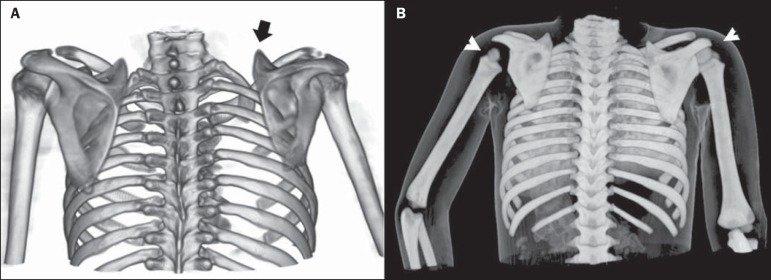
Figure 6.
Examples of three-dimensional volume rendering of computed tomography images acquired from two different patients. A: Three-dimensional reconstruction, posterior view. Female patient, 12 years old, history of high obstetric traumatic brachial plexus injury on the right. Note the elevation of the right scapula (arrow) and reduction of right scapula size. B: Posterior view. Volume rendering with skin referential of a 4-year-old male patient with obstetric brachial plexus injury on the left side. Note the elevation of the left scapula, reduced size of the humeral head (arrowheads), and positioning of the left limb (maintained in abduction and internal rotation).

