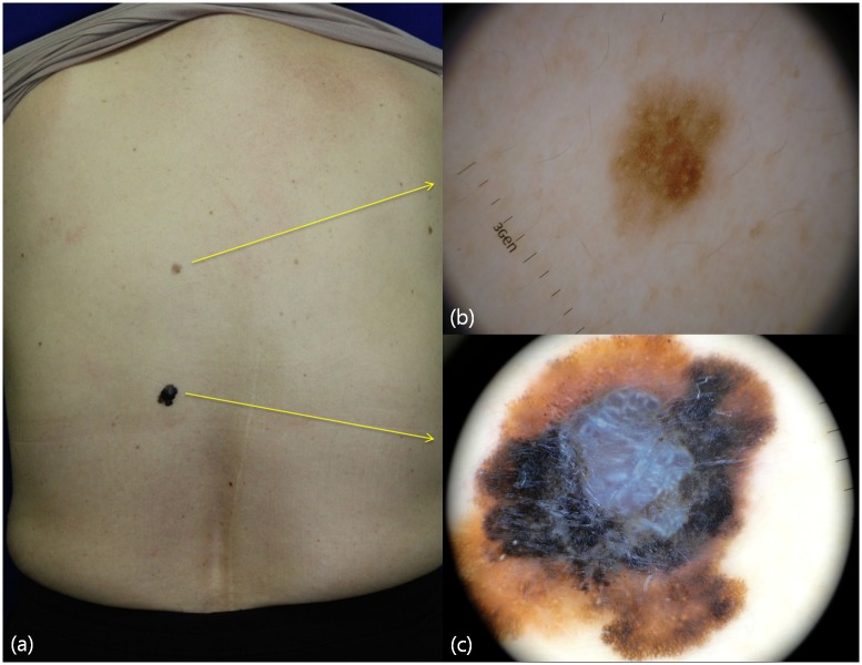
Fig 1. Clinical image and dermoscopic features of a malignant melanoma and benign nevus in a 60-year-old woman.
Compared to the clinical picture (a), dermoscopic evaluation provides detailed clues for the diagnosis of melanomas (b), dermoscopy of benign nevus showing a reticular pattern (c). Dermoscopy of malignant melanoma exhibits melanoma-associated patterns such as asymmetry, atypical pigment networks, blue-white veil, irregular blotches, irregular dots, peripheral streaks, shiny white lines, and multicolor patterns.

