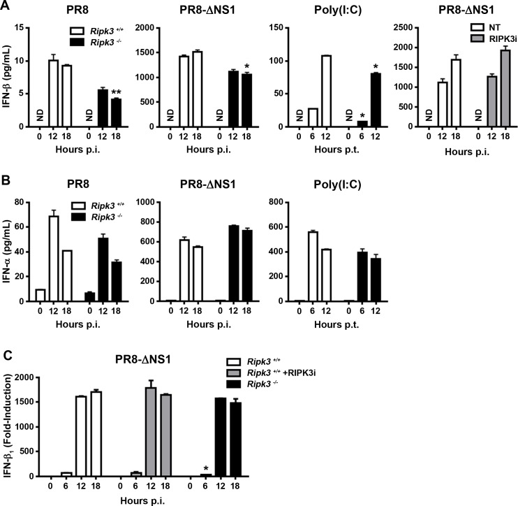
Fig 2. Modest post-transcriptional role for RIPK3 in production of IFN-β upon RLR stimulation.
(A) Ripk3+/+ or ripk3-/- MEFs were infected with WT PR8 (m.o.i. = 2), PR8-ΔNS1 (m.o.i. = 1), or transfected with virus mimetic poly(I:C) for the indicated times, and IFN-β in supernatants of cultured cells was quantified by ELISA. Ripk3+/+ MEFs were infected with PR8-ΔNS1 in the presence or absence of the RIPK3 kinase inhibitor GSK’872 (5μM), for the indicated times, and secretion of IFN-β in supernatants of cell culture was quantified by ELISA. (B) IFN-α in supernatants of cell culture was also quantified by ELISA. (C) Ripk3+/+ MEFs treated with or without RIPK3 inhibitor (GSK’872, 5μM), or ripk3-/- MEFs, were infected with PR8-ΔNS1 (m.o.i. = 1), for the indicated times, and ifnb1 mRNA levels determined from DNA microarray data output. Fold-induction was calculated using basal levels (time = 0) of each condition as baseline, which was normalized to 1. Error bars represent mean +/- S.D. NS = not statistically significant; ** p <0.005; * p <0.05.

