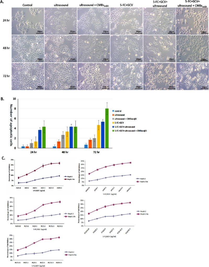
Fig 6. Effect of CMBsαvβ3in 5-FC / GCV—induced anti-proliferation in HepG2 cells.
A. HepG2 cells were treated with ultrasound alone, ultrasound + CMBsαvβ3,5-FC / GCV, 5-FC / GCV + ultrasound, or 5-FC / GCV + ultrasound + CMBsαvβ3for 24, 48 and 72 hours. Untreated HepG2 cells were served as control. Featured apoptotic cell death was observed under optical microscope (Magnitude×40). B. Numbers of apoptotic cells in each treatment group were counted for five different visual fields (mean ± SD of three experiments; *p < 0.05). C. The effect of CMBsαvβ3in 5-FC / GCV—induced anti-proliferation in HepG2 cells was measured by MTT assay. 5-FC / GCV with ultrasound plus CMBs was served as control (mean ± SD of three experiments; *p < 0.05).

