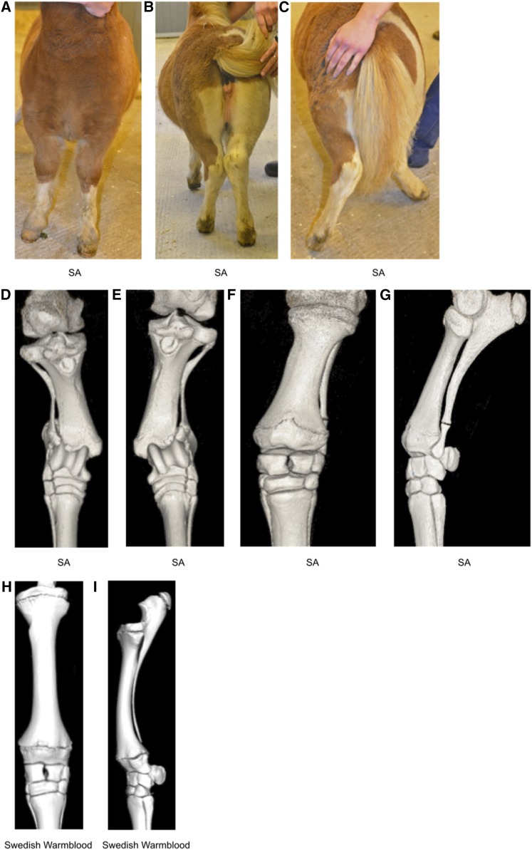
Figure 1.
Limbs of a 16-wk-old Shetland pony with skeletal atavism. (A) View from the front when standing square, (B) caudal view when standing, and (C) caudal view at walk. Complete fibulas and ulnas cause instability in the tarsocrural and antebrachiocarpal joints, respectively; angular limb deformities become more severe at walk. (D–G) Computed tomography scans of the 16-wk-old Shetland pony’s gaskin and forearm. Dorsal views of tibia and complete fibula, right (D) and left (E) hind limbs. (F) Dorsal and (G) lateral views of left front limb radius and complete ulna. (H) Computed tomography scans showing dorsal and (I) lateral views of normally developed radius and ulna, with the ulna about to be fused to the radius, of a 16-wk-old nonatavistic Swedish Warmblood foal.

