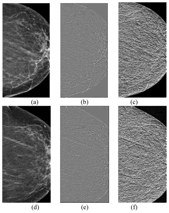
Fig. 2.
An example of a positive case in which cancer was detected on the “current” mammogram of left breast. It includes central regions extracted from the “prior #1” mammograms of the left (a) and right breast (d); WLD differential excitation images of the left (b) and right breast (e); and WLD gradient orientation images of the left (c) and right breast (f).

