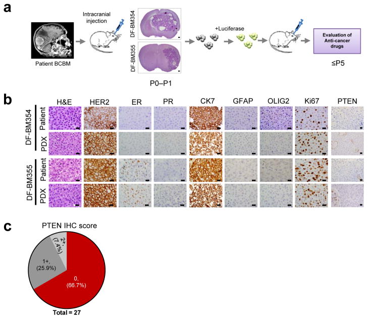
Figure 1. Establishment of orthotopic HER2-positive BCBM PDXs.
(a) Schematic depicting the process of generating orthotopic PDX BCBM models for use in preclinical studies. Fresh brain metastatic tissues from patients with BCBM were grafted directly into the brains of female SCID mice. The xenografts in the brain were explanted, dissociated and transduced with a luciferase gene, and then re-injected into new cohorts of mice. P0, primary graft; P1–P5, passage number in mice. DF-BM: Dana-Farber Brain Metastases samples. (b) Representative histologic and immunophenotypic analyses of two patient surgical biopsies and corresponding PDXs. (Scale bars = 25 μm). (c) Compiled result of PTEN immunohistochemistry performed on 27 human HER2-positive BCBM samples. 0, no staining in > 90% of tumor cells; 1+, weak staining in > 75% of tumor cells; 2+, strong staining in > 75% of tumor cells.

