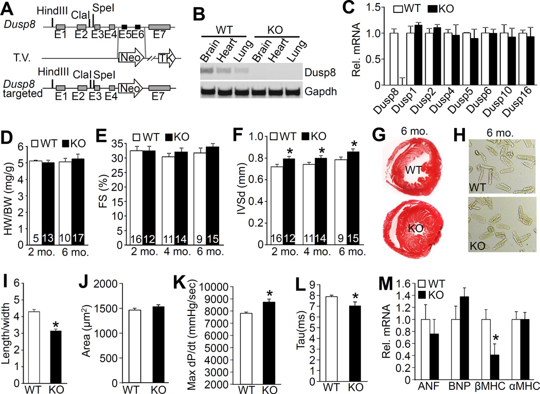
Figure 2. Gene targeting of Dusp8 and resulting cardiac phenotype.
A, Schematic of the Dusp8 genetic locus and the targeting vector (T.V.) used to create Dusp8 gene-deleted embryonic stem cells, then mice. Restriction enzyme sites and exons are shown, and the neomycin (Neo) resistance cassette in the T.V. B, RT-PCR analysis of Dusp8 mRNA in the brain, heart and lung of 2 month-old WT versus Dusp8 KO mice. Gapdh was used as PCR control. C, Real-time PCR analysis of expression of multiple Dusp mRNAs in hearts of 2 month-old Dusp8 WT or KO mice. N=3 individual samples. D, Heart weight (HW) normalized to body weight (BW) in Dusp8 WT and KO mice at the indicated ages. Number of mice used is shown in the bars. E-F, Echocardiographic assessment of FS and interventricular septal thickness in diastole (IVSd) in Dusp8 WT and KO mice at the indicated ages. *p<0.05 vs WT. Number of mice used is shown in the bars. G, Representative Masson trichrome-stained histological sections from the hearts of Dusp8 WT and KO mice at 6 months of age. Magnification is 40x total. H, Representative microscopic phase-contrast images of adult cardiac myocytes isolated from 6 month-old Dusp8 WT and KO mice. Magnification is 200x total. I, Quantification of length/width ratio of adult cardiac myocytes isolated from Dusp8 WT and KO mice at 6 months of age as shown in panel “H”. A total of 248 myocytes were analyzed for each group. *p<0.05 vs WT. J, Analysis of the surface area of adult myocytes isolated from 6 month-old Dusp8 WT and KO mice as shown in panel “H”. A total of 248 myocytes were analyzed for each group. K and L, Invasive hemodynamic measurement of (K) cardiac contractility at baseline (load-dependent) as maximum rate of pressure change in the left ventricle over time (dP/dt max) or (L) time constant for isovolumetric relaxation (Tau) showing the exponential decay of ventricular pressure during isovolumic relaxation in Dusp8 WT and KO mice. N=3 for each group. *p<0.05 vs WT. M, Real-time PCR analysis for mRNA levels of atrial natriuretic factor (ANF), b-type natriuretic peptide (BNP), β-myosin heavy chain (βMHC), and α-myosin heavy chain (αMHC) in 2 month-old Dusp8 WT or KO mice. N=4 for each group. *p<0.05 vs WT.

