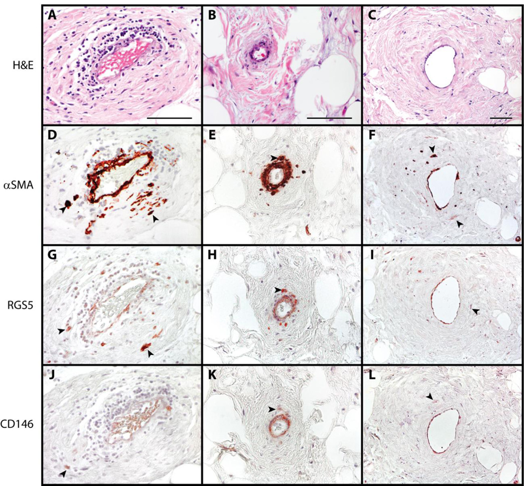
Fig. 2. Pericytic markers in liposarcoma / atypical lipomatous tumor.
(A-C) H&E appearance. (D-F) α Smooth Muscle Actin (αSMA) immunohistochemical staining. (G-I) RGS5 immunohistochemical staining. (J-L) CD146 immunohistochemical staining. Black arrowheads indicate atypical enlarged stromal cells with positive staining. Black scale bar: 100 µm.

