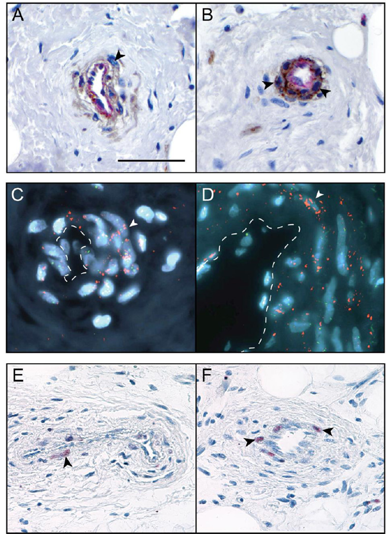
Fig. 4. Tumor cell origin of perivascular cells in liposarcoma / atypical lipomatous tumor.
(A,B) Dual immunohistochemical staining for CD31 (endothelial marker, appearing red) and CD146 (pericyte marker, appearing brown). Please note that CD146 is known to also highlight endothelial cells. (C,D) MDM2 amplification by Fluorescence In Situ Hybridization. MDM2 appears red, CEP 12 appears green, and DAPI nuclear counter stain appears blue. White dashed lines highlight vascular lumen. (E,F) Nuclear MDM2 immunohistochemical staining. Black and white arrowheads indicate atypical enlarged stromal cells with positive staining. Black scale bar: 100 µm.

