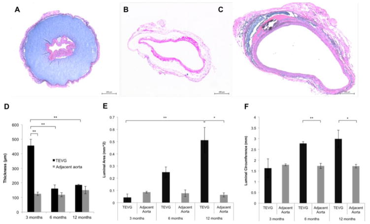
Figure 3.
H&E staining of TEVGs retrieved at 3 months (A), 6 months (B), and 12 months (C) after implantation. (D) Quantification of electrospun graft wall thickness. Graft dilatation was confirmed by quantification of luminal area (E) and luminal circumference (F) from histological images. Values are mean ± SEM. *p < 0.05, **p < 0.005. No significant dilatation of the PCL sheath was observed throughout the 12 month long implantation period (data not shown).

