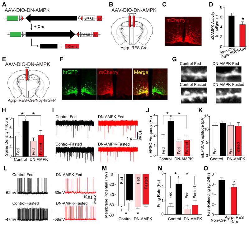
Figure 2. AMPK is required for fasting-induced synaptic plasticity in AgRP neurons.
(A–D) Schematics of dominant negative AAV-DIO-DN-AMPK (A) and stereotaxic injection (B), immunofluorescence of mCherry (C), and arcuate α2AMPK kinase activity from ad libitum fed mice (D) (n=8).
(E–N) Following unilateral injection of AAV-DIO-DN-AMPK (E), immunofluorescence (F), examples and summary of dendritic spines (G and H), mEPSCs (I–K), and firing properties (L–N) are shown (nfed=9 and nfasted=11 neurons from 3 mice per group) in fed or fasted mice.
(O) Following bilateral injection of AAV-DIO-DN-AMPK, food eaten following 24-hr fasting (n=8).
Data are mean ± SEM and * indicates p<0.05 with unpaired two-tailed student’s t-test (D and O) and with unpaired one-way ANOVA test (H, J, K, M, and N).

