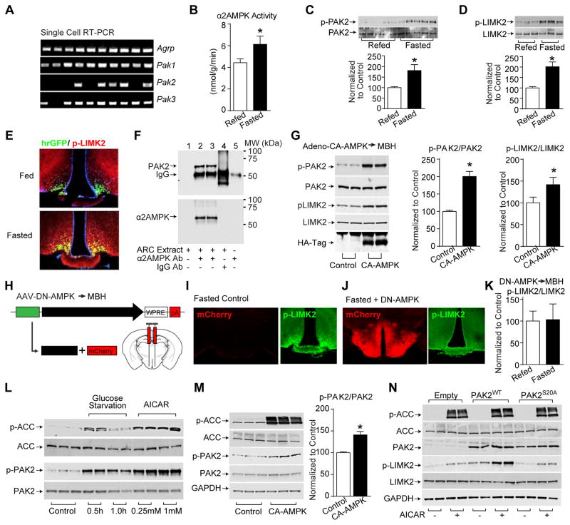
Figure 3. AMPK phosphorylates and stimulates PAK signaling.
(A) Single cell RT-PCR in AgRP neurons
(B–D) Arcuate α2AMPK activity (B) and total and phosphorylated PAK2 (Ser20) (C) and LIMK2 (Thr505) (D) in arcuate lysates from fasted and 6-hr refed wildtype mice (nrefed=9 and nfasted=8).
(E) Immunofluorescence of arcuate p-Thr505LIMK2 from fed and 24-hr fasted Npy-hrGFP mice.
(F) Immunoprecipitation of PAK2 and α2AMPK from arcuate lysates of fed wildtype mice.
(G) Phosphorylation of PAK2 (Ser20) and LIMK2 (Thr505) in the arcuate of fed wildtype mice following bilateral injection of HAtag-CA-AMPK adenovirus (n=5).
(H–K) Schematics of cre-independent AAV-DN-AMPK and stereotaxic injection into mediobasal hypothalamus (MBH) (H), immunofluorescence of mCherry (red) and p-Thr505LIMK2 (green) from fasted non-viral infected control mice (I) and AAV-DN-AMPK injected mice (J), and the ratio of total and phosphorylated LIMK2 (Thr505) in the arcuate lysates as detected with western blot from fasted and 6-hr refed mice following AAV-DN-AMPK injection (K)(n=8).
(L–N) Total and phosphorylated ACC (Ser79), PAK2 (Ser20) and LIMK2 (Thr505) in GT1-7 cells following glucose starvation or AICAR treatment (L), or transfection of CA-AMPK (n=9) (M), or transfection of PAK2WT and PAK2S20A mutants with 1 mM AICAR treatment (N). Proteins are normalized to GAPDH.
Data are mean ± SEM and * indicates p<0.05 with unpaired two-tailed student’s t-test.

