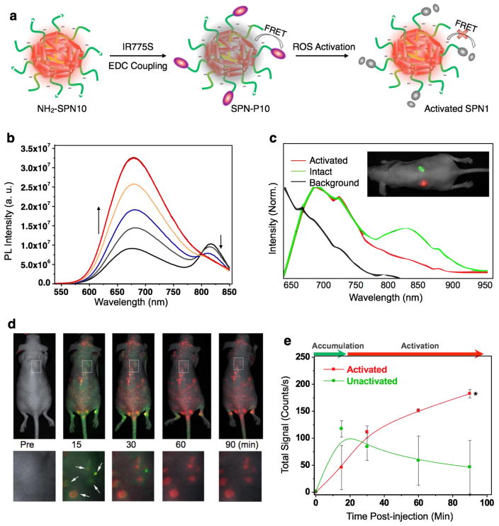
Fig. 4.
(a) Schematic of the preparation and RONS sensing of SPN-P10. (b) Fluorescence spectra of SPN-P10 in PBS (30 mM, pH = 7.4) in the absence or presence of ONOO− with concentrations ranging from 0 to 0.5 μM at intervals of 0.1 μM. (c) Unactivated (green) and preactivated SPN-P10 (red) were injected subcutaneously into the back of a nude mouse, followed by fluorescence hyperspectral imaging to record the in vivo spectra of the activated (red) and unactivated probes (green), as well as autofluorescence (black). (d) Imaging RONS with SPN-P10 in mice with spontaneous systemic C. bovis bacterial infection. Overlaid images of activated (red) and unactivated (green) SPN-P10 following i.v. administration to mice with spontaneous bacterial infections (n=4). Enlargements of the regions indicated by dashed white boxes are given below each corresponding image. White arrows indicate localized regions of bacterial infection. (d) Quantification of activated (red) and non-activated (green) SPN-P10 fluorescence over time. †Significantly different change in fluorescence between unactivated and activated nanoprobe (p<0.05). Reproduced from ref. 69.

