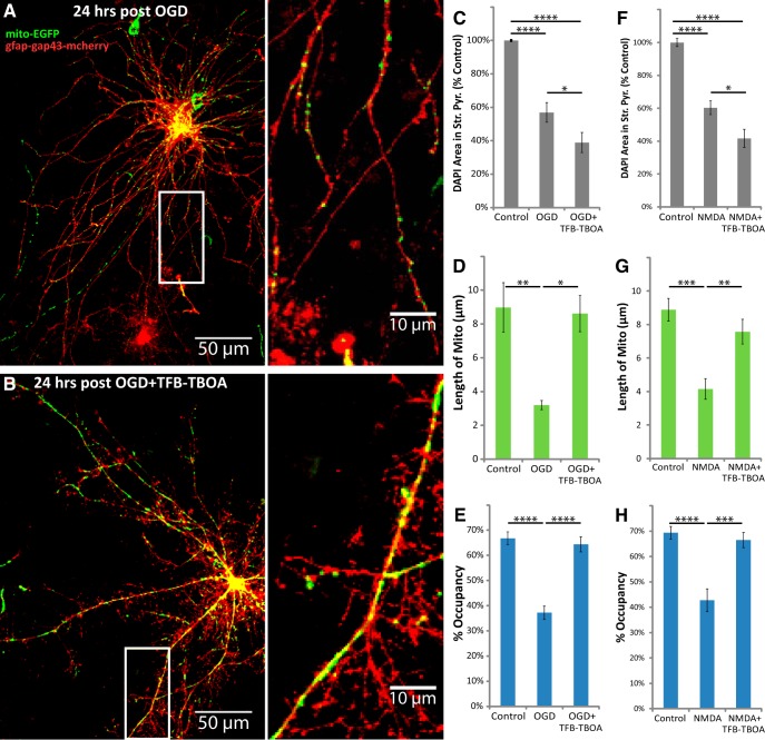
Figure 6.
TFB-TBOA increased neuronal loss, but blocked the loss of astrocytic mitochondria after OGD or NMDA injury. Hippocampal slice cultures with astrocytes expressing mitochondrially targeted EGFP (mito-EGFP, green) and plasma-membrane-targeted mcherry (gfap-gap43-mcherry, red) were fixed in 4% paraformaldehyde 24 h after a 30 min OGD, NMDA (100 μm), or control treatment. OGD and NMDA slices were treated 30 min before insult, during the 30 min insult, and during the 24 h recovery period either with no drug or with 3 μm TFB-TBOA. A, B, Representative images of fluorescently labeled mitochondria (green) and plasma membrane (red) in astrocytes fixed 24 h after OGD (A) or OGD in the presence of TFB-TBOA (B). C–H, Mean cell density in stratum pyramidale, mitochondrial length in processes (micrometers), and percentage occupancy of processes by mitochondria 24 h after 30 min OGD (C–E) or NMDA (F–H) insult with or without TFB-TBOA. All groups were included in ≥3 experiments, with slice cultures prepared from ≥3 separate animals. n = 8–12 slices/group (2–4 slices/group/experiment, 2 astrocytes/slice, 3 processes/astrocyte). Error bars indicate SEM. *p < 0.05, **p < 0.01, ***p < 0.001, ****p < 0.0001 compared by one-way ANOVA with Bonferroni's correction for multiple comparisons. Insets provide magnified views of mitochondria in astrocytic processes.

