Figure 9.
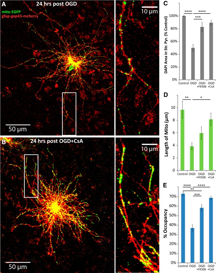
CsA or FK506 blocked the loss of astrocytic mitochondria after OGD. Hippocampal slice cultures with astrocytes expressing mitochondrially targeted EGFP (mito-EGFP, green) and plasma-membrane-targeted mcherry (gfap-gap43-mcherry, red) were fixed in 4% paraformaldehyde 24 h after 30 min OGD or control treatment. OGD slices were treated 30 min before insult, during the 30 min insult, and during the 24 h recovery period either with no drug, with 10 μm CsA, or with 10 μm FK506. A, B, Representative images of fluorescently labeled mitochondria (green) and plasma membrane (red) in astrocytes fixed 24 h after OGD (A) or 24 h after OGD in the presence of CsA (B). Insets provide magnified views of mitochondria in astrocytic processes. C–E, Effects of CsA or FK506 on mean cell density in stratum pyramidale (C), mitochondrial length in processes (micrometers; D), and percentage occupancy of processes by mitochondria (E) 24 h after 30 min OGD. All groups were included in ≥3 experiments, with slice cultures prepared from ≥3 separate animals. n = 6 slices/group (2 slices/group/experiment, 2 astrocytes/slice, 3 processes/astrocyte). Error bars indicate SEM. *p < 0.05, **p < 0.01, ***p < 0.001, ****p < 0.0001 compared by one-way ANOVA with Bonferroni's correction for multiple comparisons.

