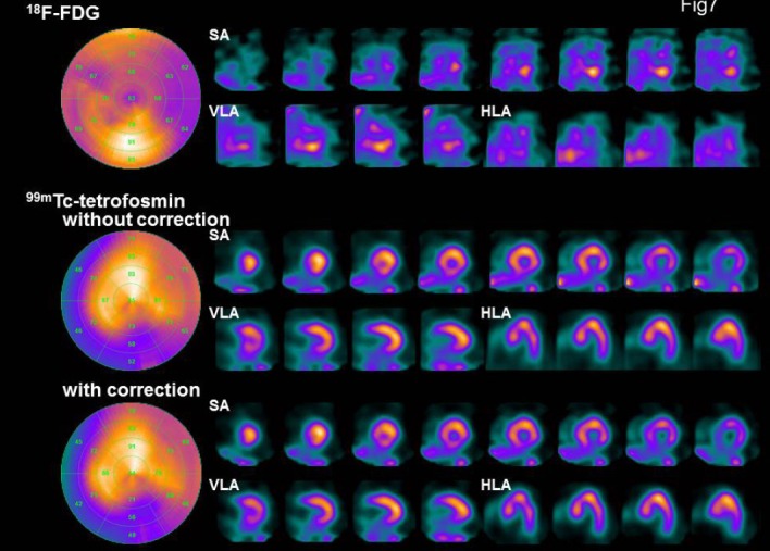
Figure 7.

Bull’s eye map; SA: short axis, VLA: vertical long axis, and HLA: horizontal long axis in the evaluated patient. 99mTc-tetrofosmin SPECT images were obtained before and after DEW crosstalk correction. An 81-year-old male was admitted to our center with chest pain. His risk factors for coronary artery disease included a prior history of hypertension and a family history of ischemic heart disease. The electrocardiogram showed ST-segment elevation in II, III, and AVF. The patient underwent selective left and right coronary artery angiography. The left coronary angiography showed moderate stenosis in segment VI. The right coronary angiography revealed 90% stenosis in segment I. He underwent stent placement in segment I. Seven days after stenting, simultaneous 18F-FDG and 99mTc-tetrofosmin SPECT was performed
