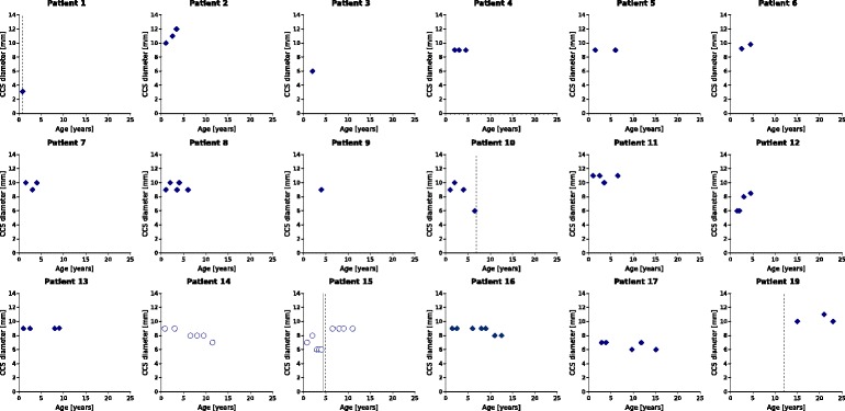
Fig. 3.

Longitudinal MRI assessments of CCJ diameters. The shortest anterior-to-posterior diameters of the CCJ (vertebrae C1-C3) were assessed on sagittal T2-weighted MRI scans and are shown for each patient separately. The two patients with graft failure (patient 14 and 15) are presented as circles. Dashed straight lines indicate the time of surgical intervention. The straight line represents the start of ERT
