
Fig. 5.
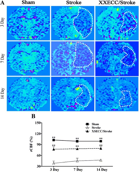
CBF images on the 3rd, 7th and 14th days after pMCAO. a Representative CBF images of the Sham, the Stroke and the XSECC/Stroke groups. The low cerebral flow blood as indicated by the white dotted line. b rCBF on the 3rd, 7th and 14th days after pMCAO. Values are means ± SEM. **P < 0.01vs the Stroke group