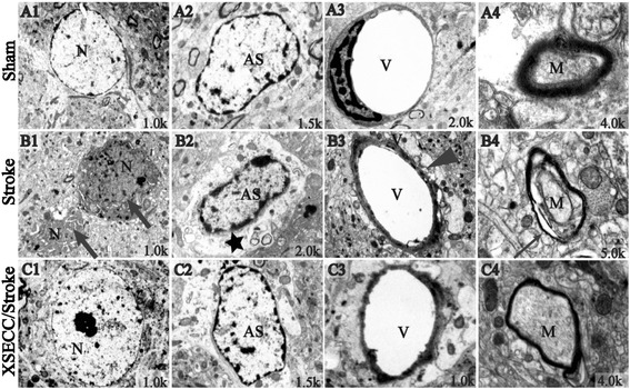
Fig. 7.

Ultrastructure of the NVU in parietal cortex on the 14th day after pMCAO. Representative images of ultrastructure in (A1-A4) the Sham group, (B1-B4) the Stroke group and (C1-C4) the XSECC/Stroke group. Normal appearance of (A1) neurons and (A2) astrocytes; (A3) the regular capillary; (A4) integral myelin. (B1) Degenerative and necrotic neurons as indicated by broad arrows; (B2) Swollen astrocyte foot processes as indicated by black star; (B3) edema around the capillaries as indicated by black arrow heads and (B4) disrupted Myelin sheath as indicated by narrow arrow. n = 2 for each group. N: neurons; AS: astrocytes; V: vascular. M: myelin sheath
