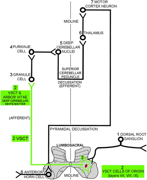
Fig. 6.

Schematic of the anatomical pathways that involve the VSCT. Afferent input from the peripheral nervous system via the (1) dorsal root ganglia synapse on the cells of origins (neuronal cell bodies) of the (2) VSCT, which are contained within laminae VII, VIII, and IX of lumbosacral levels of the spinal gray matter. Axons of the (2) VSCT, which are preferentially damaged by the anti-hnRNP A1-M9 antibodies (shown in green), immediately cross the mid-line of the spinal cord. The (2) VSCT then passes through the brainstem where it enters the cerebellum parallel to the superior cerebellar peduncle. (2) VSCT axons then pass through the deep cerebellar white matter to synapse on (3) granule cells. (3) Granule cells synapse on (4) Purkinje cells, which in turn, synapse in (5) the deep cerebellar nuclei. The (5) deep cerebellar nuclei send efferent axons via the superior cerebellar peduncle to the (6) thalamus, which relays information to the (7) motor cortex, which in turns sends axons that synapse on (8) anterior horn cells in the ventral gray of the spinal cord. The (2) VSCT (green) showed neurodegeneration from the spinal cord up through to its distal axons in the deep white matter of the cerebellum, where it was preferentially affected by the anti-hnRNP A1-M9 antibodies consistent with “dying back” axonal degeneration
