Fig. 6.

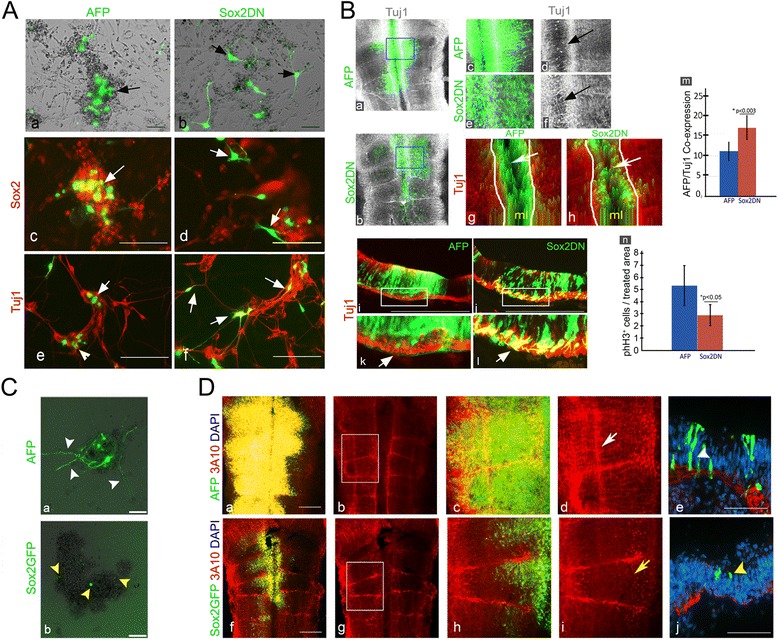
Sox2 regulates neural differentiation within the developing hindbrain. A Representative primary cultures prepared from 18HH hindbrains electroporated with control AFP (n = 12) or Sox2DN (n = 12) plasmids (green). Bright-field (a,b) and immunostaining with Sox2 (c,d) or Tuj1 (e,f). Arrows indicate electroporated cells. B Representative flat-mounts (a–f) (n = 10 in each treatment) and transverse sections (i–l) (n = 5 in each treatment) of 18HH hindbrains electroporated with AFP/Sox2DN (green) and stained for Tuj1 (grey/red). High magnification views of boxed areas in (a,b,i,j) are shown in c,d,k for AFP and in e,f,l for Sox2DN. Arrows in (d,f) indicate Tuj1-expression in the midline and in (k,l) indicate Tuj1+ fibers in the mantle zone. 2.5D plots obtained from flat-mounted hindbrains (g,h). Arrows indicate the midline. Flow cytometry quantification of hindbrains expressing AFP+ or Sox2DN+ cells that express Tuj1 (m). Quantification of phH3+ cells per area in AFP or SoxDN-treated hindbrains (n). C Primary cultures prepared from 18HH hindbrains electroporated with control AFP or Sox2GFP plasmids (green). Arrowheads indicate electroporated cells. D Representative flat-mounted views of AFP (n = 15) or Sox2GFP (n = 17) electroporated hindbrains stained for 3A10 (red) (a–d,f–i). Images in (a,c,f,h) show also AFP/GFP expressing cells. Enlargement of boxed regions in (b,g) is shown in (c,d,h,i). White and yellow arrows indicate typical and reduced 3A10+ fibers in rhombomeres, respectively. (e,j) Transverse sections of electroporated hindbrains. White arrowhead denotes AFP+ cell near the MZ; yellow arrowhead denotes Sox2GFP+ cell in the VZ. ml = midline. Scale bars = 100 μm
