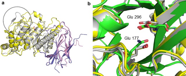
Fig. 4.

Modeled structures of THSAbf. a The overall two domain structure composed of a (β/α)8 folded catalytic domain and a C-terminal domain displaying β-sandwich architecture. Two structural conformers of THSAbf (open gray and violet, closed yellow and pink) are superposed and the β2α2 loop in two different positions is encircled. b Zoom on the catalytic site of the two modeled THSAbf conformers (gray and yellow) and that of GsAbf (1QW8, green). The side chains of the two catalytic residues (Glu 177 and 296 in THSAbf) are shown as sticks. (Figure prepared using PyMOL™ Molecular Graphics System, Version 1.7.2.1)
