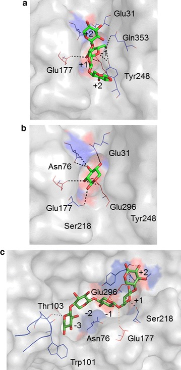
Fig. 5.

Docking of various ligands into the active site of THSAbf. a XA3XX, from 2VRQ, b a single β-d-xylosyl moiety from 1QW8 and c xylopentaose (X5). Sugar moieties in the oligosaccharides are numbered according to their position with regard to the putative scissile bond, with most negative number designating the non-reducing moiety. The side chains of amino acids that might form polar contacts (dotted lines) with the sugar ligands are shown (blue lines) and the active site residues Glu177 and Glu296 are highlighted using red lines and in c the β2α2 loop is shown as a blue ribbon. (Figure prepared using PyMOL™ Molecular Graphics System, Version 1.7.2.1)
