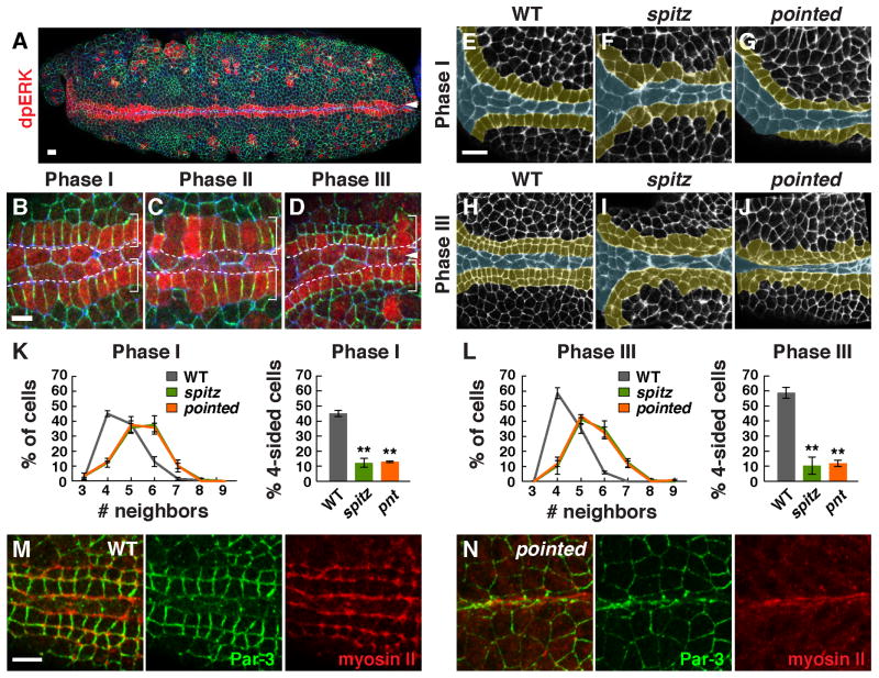
Figure 5. EGF receptor signaling is required for square grid formation.
(A–D) MAP kinase activity (indicated by dpERK signal) was increased in the first row of square grid cells adjacent to the midline in phase I (A and B). DpERK signal decreased in lateral daughter cells after division (C,D). DpERK (red), Par-3 (green), E-cadherin (blue). (E–J) Cell alignment and apicobasal cell elongation were disrupted in spitz (F,I) and pointed (G,J) mutants compared to wild type (E,H) (F-actin, white; square grid region, yellow). (K, L) The percentage of four-sided cells was significantly lower in spitz (p = 0.0001 in late Phase I, 0.0002 in Phase III) and pointed mutants (p ≤ 0.0001 in late Phase I and Phase III) compared to wild type (unpaired t test, n = 70–145 cells in 4 embryos/genotype in Phase I and 145–398 cells in 4–7 embryos/genotype in Phase III). (M,N) Myosin II localization parallel to the midline did not occur between adjacent rows of square grid cells in pointed mutants, but myosin accumulation at interfaces contacting the midline occurred normally. Bars, 10 μm (A, E–J), 5 μm (B–D,M,N).

