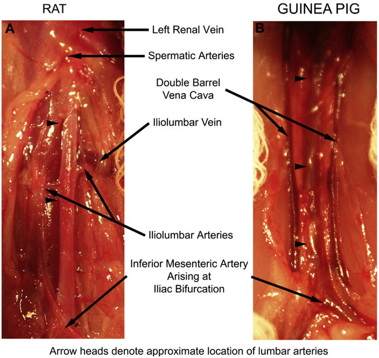
Fig 5.

Pertinent aortic anatomy of the (A) rat and (B) guinea pig. Limits of the dissection include the renal vein and spermatic arteries cranially and the inferior mesenteric arteries and bifurcation of the aorta into the iliac arteries caudally. The iliolumbar vein marks the location of the iliolumbar artery and the approximate locations of the five lumbar arteries that arise from the dorsal surface of the aorta. Branches ligated include the paired iliolumbar and proximal lumbar arteries in the rat and the lumbar arteries in the guinea pig. Note the common occurrence of a double-barrel inferior vena cava in the guinea pig. The arrowheads denote the approximate location of the lumbar arteries ligated in the (A) rat and (B) guinea pig.
