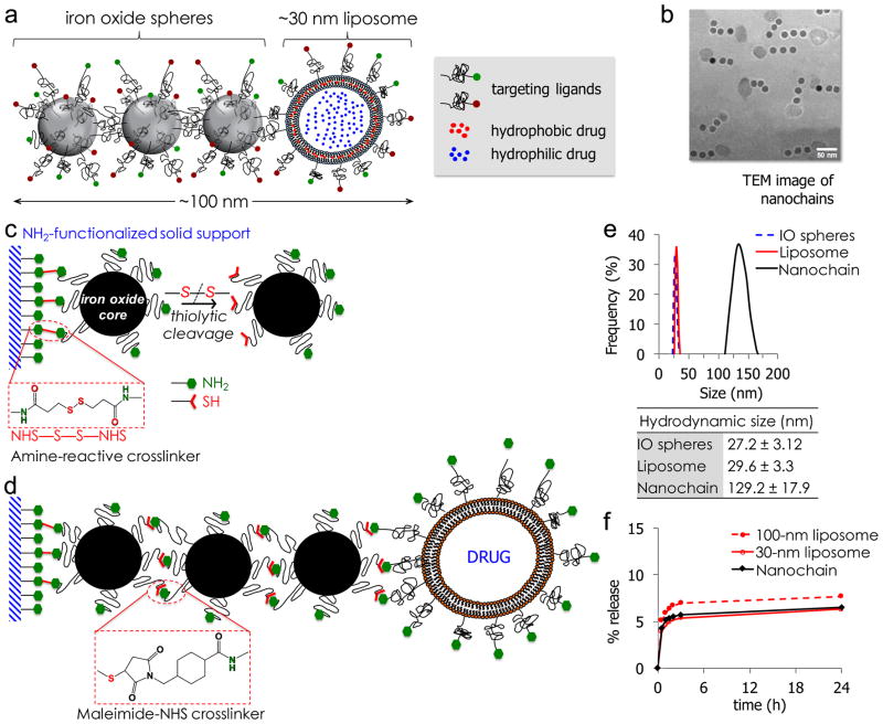
Figure 1.
(a) Schematic of a linear nanochain particle composed of three IO nanosphere and one drug-loaded liposome. (b) TEM image of nanochain particles (reprinted with permission after partial modifications from ref. 11). (c) Reaction scheme of the controlled assembly of nanochains using solid-phase chemistry. In the first step, chemical bifunctionality on the surface of parent IO nanospheres is topologically controlled resulting in nanospheres with two faces, one displaying only amines and the other only thiols. (d) In the second step, the two unique faces on the parent nanosphere serve as fittings to chemically assemble them into nanochains. (e) Size distribution of nanochain particles and their parent nanospheres obtained by DLS (data presented as mean ± s.d.). (f) Comparison of in vitro blood plasma stability of nanochains to 30-nm and 100-nm liposomal DOX. In a typical leakage procedure, 1 mL of formulation was placed in dialysis tubing with 100k MWCO and dialyzed against blood plasma at 37°C. (reprinted with permission after partial modifications from ref. 12).

