Fig. 4.
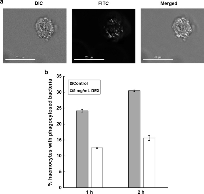
a Representative image of a single G. mellonella haemocyte after 1 h incubation at 37 °C with inactivated FITC-labelled E. coli K12. Images are shown with differential interference contrast (DIC), filters for FITC (excitation 490 nm/emission 528 nm) and a merged DIC/FITC image. The merged image represents a central 0.2 μM optical slice through the haemocyte allowing precise counting of internalised bacteria. Haemocytes and fluorescent E. coli were viewed at 40× magnification. b Effect of a 1- or 2 h incubation at 37 °C with dexamethasone 21-phosphate (DEX; 5 mg/mL) on the phagocytosis of inactivated fluorescein (FITC)-labelled E. coli K12 by purified haemocytes from G. mellonella larvae. Haemocytes and fluorescent E. coli were viewed using differential interference contrast or fluorescence microscopy at 40× magnification, and bacteria inside the haemocytes were counted. For each time point tested, phagocytosed E. coli from at least 30 haemocytes, from at least two replicate experiments, were counted per slide. ±SE bars are drawn for each treatment group

