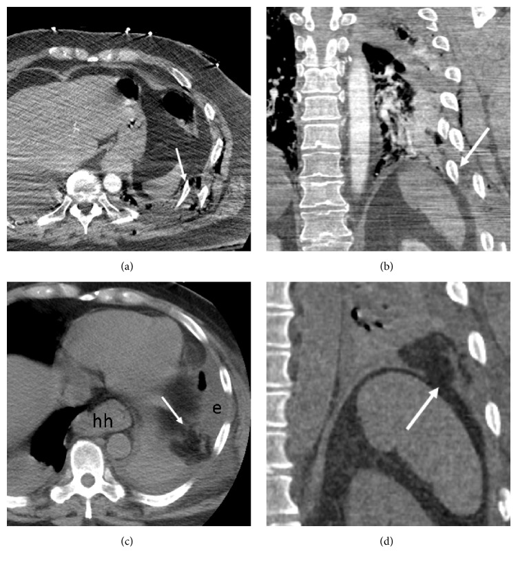
Figure 2.
Fractured rib sign in a 64-year-old man after blunt trauma. (a, b) Contrast-enhanced trauma chest MDCT (axial and coronal planes) shows displaced left lower rib fractures abutting left diaphragm (arrows). The diaphragm appears intact. A small hiatal hernia is present (hh). (c, d) Repeat MDCT 4 days later (axial and coronal planes) shows new herniated omental fat (arrows) within left hemithorax due to subacute diaphragm rupture. Note discontinuity of the diaphragm (d). The large left effusion (e) is new.

