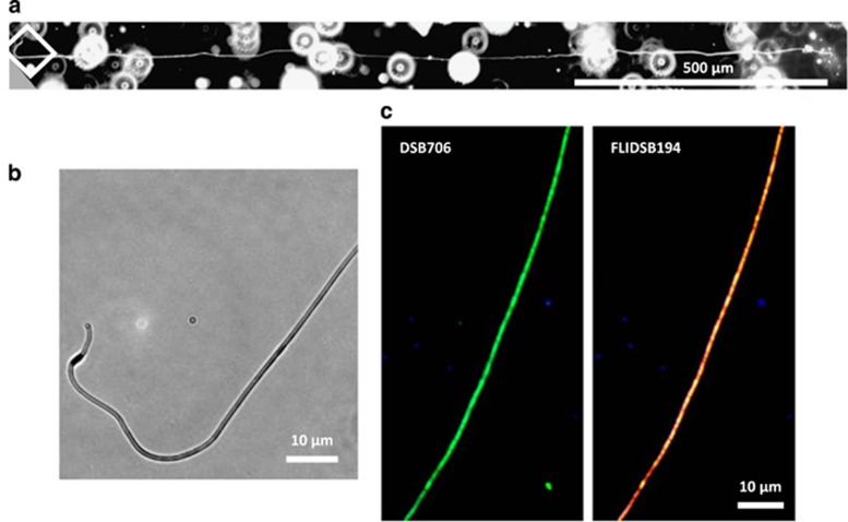
Figure 2.
(a) Microscopic image of a filament fragment of ~1.7 mm length. Three pictures were merged to cover the full length of the fragment. The white square indicates the part of the filament shown in higher magnification in (b). (c) Micrographs of filaments stained with FISH probes specific for the family Desulfobulbaceae (DSB706; 6-FAM labeled) and for groundwater cable bacteria (FLIDSB194, Cy3 labeled). Each image is presented as an overlay of two pictures taken with filters for specific probe fluorescence and 4',6-diamidino-2-phenylindole for counterstaining.

