
FIGURE 6.
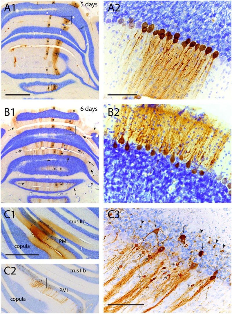
Failure of transneuronal infection from Purkinje cells to molecular layer interneurons and granule cells. (A1,2) First appearance of labeled neurons 5 days after injection of anterior tibial muscle. Strips of infected Purkinje cells are seen in the lateral A-zone and within the B-zone of the cerebellar cortex (marked by asterisks). Boxed area in (A1) is enlarged in (A2). Within the B-zone, as expected, no other neurons than Purkinje cells are labeled. (B1,2) Again A- and B-zone labeling (asterisks) in a similar experiment but now with a survival time of 6 days, resulting in many additional RABV-labeled strips of Purkinje cells. However, careful examination of the B-zone did not show any infected stellate, basket or granule cells. (C1–3) Injection site (encircled) of RABV/CTb mixture injected directly in the cerebellar cortex. (C1) CTb immunohistochemistry, (C2) RABV immunohistochemistry. (C3) Detail of boxed area shown in (C2). Note many labeled types of cerebellar cortical interneurons (e.g., stellate/basket cells: double headed arrows; Golgi cells, arrows; granule cells: arrowheads). Scale bars equal 1 mm (A1–C1,C2) and 100 μm (A2,B2,C3).