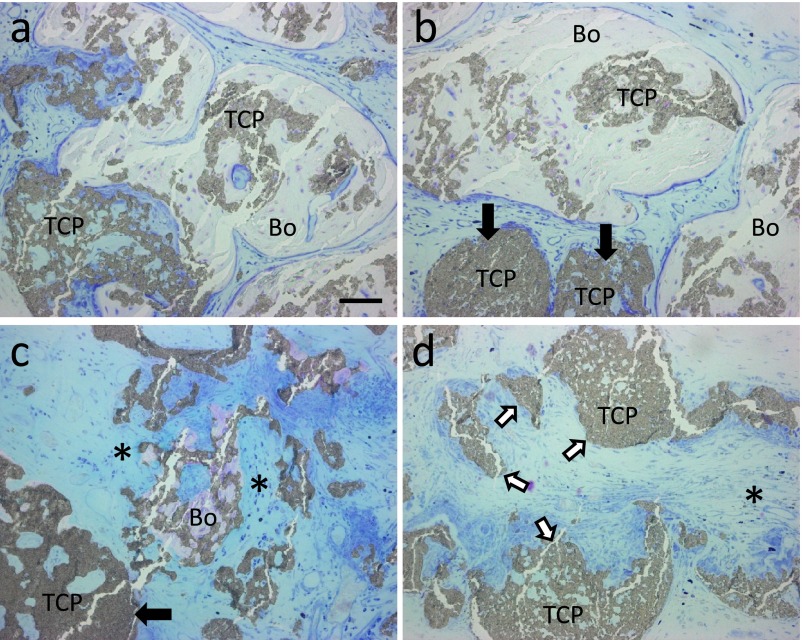
Fig. 4.
Histological views of specimens implanted with β-TCP granules with BMMCs. Sections were stained with Giemsa solution. (a, b) Residual β-TCP granules (TCP) were surrounded by a large amount of bone (Bo). (c) Partially absorbed β-TCP fragments were adjacent to a small amount of bone tissue and surrounded by connective tissue (*). Black arrows indicate residual β-TCP granules that were poorly degraded. (d) The region without osteogenesis, which includes dissociated β-TCP granules (white arrows) and β-TCP granules with large deficiencies, was filled with connective tissue (*). Bar=50 μm.

