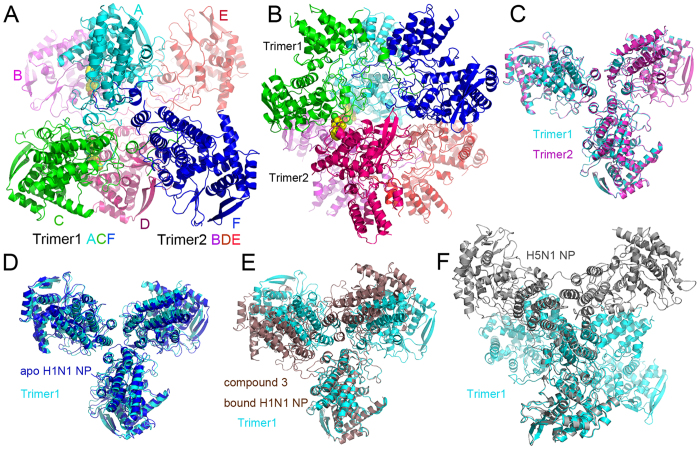
Figure 1. H1N1 NP-nucleozin complex in the asymmetric unit and comparison with other NP structures.
There were six NP molecules (molecules A–F) in the asymmetric unit and they were coloured in cyan (A), magenta (B) green (C), pink (D), red (E) and blue (F) in panels A and B. These six molecules formed dimer of trimers. Trimer 1 was composed of chains A, C and F while trimer 2 was composed of chains B, D and E. Two nucleozin molecules were found between chains A and B, C and D, respectively. They were shown in space-filling model with yellow carbon. (A) Top view of dimer of trimers. (B) Side view of dimer of trimers. The two trimers in the asymmetric unit can be superimposed perfectly with an r.m.s.d. value of 0.28 Å (C). The trimer was superimposed to apo H1N1 NP trimer (D, blue), Compound 3 bound H1N1 NP (E, brown) and H5N1 NP (F, grey). It is obvious that the NP-nucleozin structure was very close to apo H1N1 NP, but not compound 3 bound NP nor H5N1 NP.

