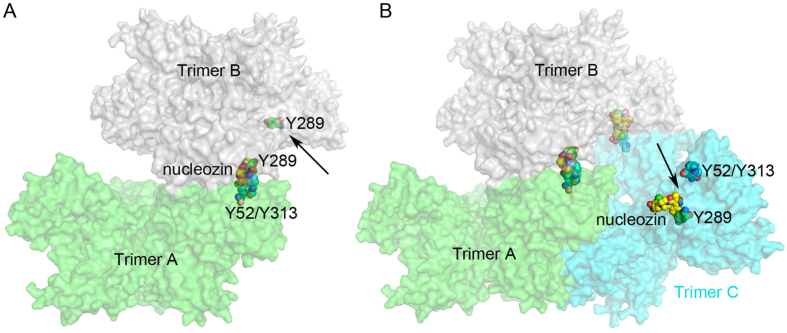
Figure 6. Model for NP-Nucleozin aggregation using the Y289/N309 and Y52/Y313 pockets.
NP trimers were shown as surface representation and colored in green (trimer A), grey (trimer B) and cyan (trimer C). Nucleozin, Y289 pocket and R382 pockets were shown as sphere models with yellow, green and cyan carbons, respectively. (A) Trimer B docked on trimer A. Unoccupied nucleozin binding pocket in trimer B was indicated by a black arrow. (B) Trimer C docked onto trimer B. There were still nucelozin binding sites available in trimer C (indicated by the black arrow).

