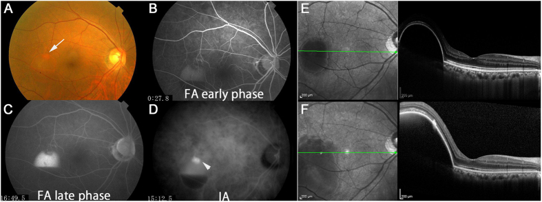
Figure 2. A non-responder as determined by fundus findings with serous pigment epithelial detachment (PED) in the treatment-naive group.
This was a 72-year-old woman with treatment-naive polypoidal choroidal vasculopathy and best-corrected visual acuity (BCVA) of 1.0 in decimal (logMAR 0) at baseline. Consistent with the findings of the fundus color photograph (A, the arrow corresponds to polyp lesion) and fluorescein (B,C, early phase and late phase respectively) and indocyanine green (D, the arrowhead shows polyp lesion) angiograms, an optical coherence tomography image showed serous PED at baseline (E). Although BCVA did not change after 7 IVA injections, serous PED worsened at month 12 (F).

