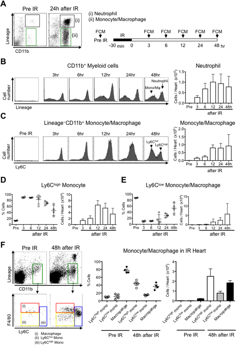
Figure 1. Ischemia-reperfusion mobilizes neutrophils and monocytes/macrophages to the heart.
(A) Flow cytometric analysis of blood neutrophils and monocytes/macrophages. Monocytes/macrophages were identified as CD11bhighLineagelowLy6Chigh/low, and neutrophils were identified as CD11bhighLineagehigh. Lineage refers to the combination of CD90, B220, CD49b, NK1.1 and Ly6G monoclonal antibodies. (B) Representative histograms from individual mice within the CD11bhigh Myeloid cells gate depict neutrophils and monocytes/macrophages in healthy hearts and within the heart at specified times after IR. Column graphs illustrate the total number of neutrophils per heart (N = 4 mice per group). (C) Representative histograms from individual mice within the monocytes/macrophages cell gate depict Ly6Chigh and Ly6Clow monocytes in healthy hearts and within the heart at specified times after IR. Column graphs illustrate the total number of monocytes/macrophages per heart (N = 4 mice per group). (D) The percentage of Ly6Chigh monocytes/macrophages and the total number of Ly6Chigh monocytes/macrophages per heart. (E) The percentage of Ly6Clow monocytes/macrophages and the total number of Ly6Clow monocytes/macrophages per heart. (F) Flow cytometry of leukocytes in the healthy hearts and hearts 48 h after IR injury. Red gate shows the population of macrophages (CD11bhighLineagelowF4/80highLy6Clow cells). The percentage and the number of macrophages, Ly6Chigh monocytes and Ly6Clow monocytes per CD11bhighLineagelow cells in IR heart. Data are expressed as the mean ± SEM (N = 4 mice). All results are average of three replicates. IR: ischemia reperfusion, FCM: flow cytometry, Mφ: macrophage, Mono: monocyte.

