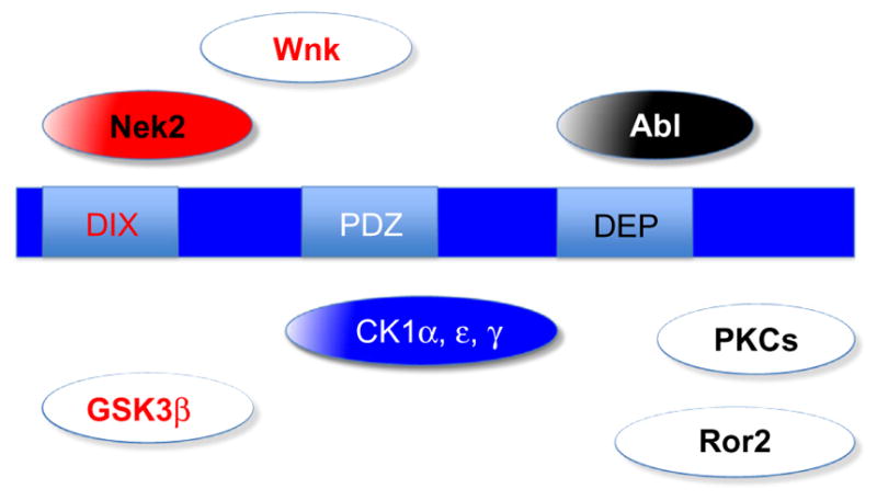
Figure 2.

Domain organization of and kinases associate with Dsh function. Color scheme is the same as in Fig. 1. Kinases that are known to directly phosphorylate Dsh are shown in pathway-specific colored solid boxes or in blue (if generally required for Dsh function, e.g., the CK1 family). Some of the more speculative kinases linked to Dsh function and phos-phorylation (but not direct phosphorylation of Dsh) are shown as white boxes with pathway-specific colored labels. Only a very limited number of kinases that have been linked to Dsh signaling are shown to reflect a conceptual (over)simplified view. See text for details.
