

Abstract
Acinic cell carcinoma arising in salivary glands is a rare tumor, accounting for 2% to 5% of the primary neoplasms of the parotid gland. When these tumors are well-differentiated, the neoplasia has innocuous aspect, due to the similarity to normal parotid tissue. This makes the diagnosis difficult. Initially the malignancy of this tumor was uncertain; however, recent studies have declared it as malignant. The female / male ratio is 3:2. The nodule usually presents as solitary and well defined shape. Several authors have used different terms to describe histomorphological patterns of these tumors. Four descriptive categories (solid, microcystic, papillary-cystic and follicular) are useful for pathologists. Here we report a case of a 49 yr old man with a left parotid nodule of 5 cm. Parotidectomy was performed at the Hospital Universitario Miguel Servet, in Zaragoza (Spain). The microscopy showed a tumor with acinic semblance, having the four morphologic patterns previously described. The morphological and immunohistochemical study was consistent with the diagnosis of acinic cell carcinoma.
Key Words: Acinic cell carcinoma, Morphologic patterns, Salivary gland
Introduction
Acinic cell carcinoma (ACC) originated in the salivary glands is a rare tumor, representing about 2% to 4% of primary neoplasms of the parotid gland (1, 2). It is formed by acinic cells describing a pattern with little stroma visible.
In well differentiated tumors, the neoplasia has innocuous appearance, because of the similarity between the normal parotid tissue normal and the neoplastic one (3), in both morphological and biochemical aspects. That can make it more difficult to diagnose.
Initially, the malignancy of this tumor was uncertain; however, more recent research, point it out as definitely malignant tumour (2, 3), with varying degrees of aggressiveness, being generally a low-grade malignant neoplasm (4). Recurrences and lymph node metastases are associated with poor prognosis.
It is located almost exclusively in the parotid gland, with a female / male ratio of 3:2, with no predilection for race and having age range from children to the elderly, with a mean of 44 yr old. It usually presents as a well-defined solitary nodule (4-6).
Various authors have used a variety of terms to describe several histomorphologic patterns and cellular features of these tumors (7). Thus, Batsakis et al. have described seven different histologic patterns for ACC: acinar-lobular, microcystic, follicular, papillary-cystic, medullary, ducto-glandular and primitive tubular (8). However, four descriptive categories (solid, microcystic, papillary-cystic and follicular) presented by Abrams et al. in 1965 have been useful to pathologists and are still applicable nowadays (6).
In the microcystic and papillary foci there are acinic cells that produce a beaded pattern or hobnail, covering cystic spaces.
The most common patterns of growth, in decreasing order, are solid feature, microcystic, papillary-cystic and follicular (6). Mostly, there is only one pattern seen in a single lesion. The combination of two or three patterns may be found, although obtaining all the four patterns in the same patient is a rare event. Solid and microcystic have been frequently associated simultaneously in previous reports (6).
Schwarz et al., described and characterized 40 cases of ACC (7). They found again the solid pattern as the most common, with 20 cases. Followed by microcystic (15 cases), and the rest of the cases having papillary-cystic, follicular, or a combination of morphological patterns.
Histological prognostic factors, such as gross invasion, desmoplasia, atypia or increased mitotic activity, are considered predictors of disease progression (4). Batsakis (8) has described a histological grading of ACC, which identifies 3 grades, I being the least infiltrative, and grade III the contrary, the most invasive.
This tumor originates as a result of neoplastic proliferation and abnormal cytodifferentiation of reserve cells or pluripotent stem cells that normally reside at the junction of acini to intercalated duct or in the intercalated duct cells itself in the mature salivary glands (4).
Due to the rarity of a case expressing the entire four main morphological types of ACC in the salivary glands, it is important to describe this neoplasm as a case report, with the aim of being well informed about the features of the tumor, to recognize easily the accurate diagnosis.
Case Report
A 49-year-old male with a tumoural lesion in the left parotid region of 5 cm consulted to Hospital Universitario Miguel Servet, in Zaragoza (Spain), and a fine needle aspiration cytology (FNAC) was performed (Fig. 1), showing abundant groups of cells with large nuclei and granular eosinophilic cytoplasm, without true acini and adipose tissue. The background was clean but bloody. It was then reported as positive for malignancy, to rule out ACC or mucoepidermoid carcinoma. Parotidectomy and left radical neck dissection were performed. Macroscopically, a tumor inside the parotid gland was found, with nodular and solid growth measuring 4.5 cm (Fig. 2). Twenty five lymph nodes were isolated.
Fig. 1.

Fine needle aspiration cytology. Papanicolaou staining (Original figure
Fig. 2:
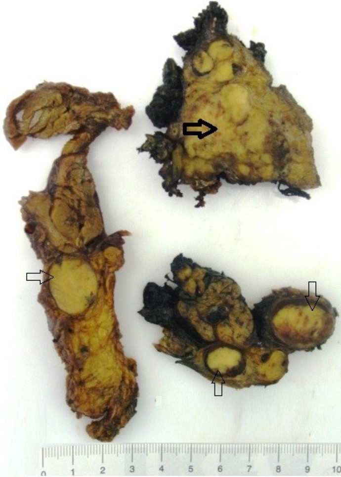
Gross specimen. The primary tumour is in the top right, with a thick arrow. Some of the metastasized lymph nodes are indicated by thin arrows
The histological study identified an epithelial neoplasia with acinic appearance. It was surprising that all four different histological patterns were seen in the same sample biopsy, the papillary-cystic pattern, follicular, solid and microcystic, with an average degree of differentiation (Fig. 3). The boundaries among the different patterns were not always neat, and two or three distinct features were found in collision (Fig. 4). Histochemistry showed cytoplasmic glycogen (PAS +, diastase-sensitive), and immunophenotype expression of CK7, CD56, CD57, IgA, CD15 and CD117/cKit (Fig.5), but not CEA, p63 or thyroglobulin. Besides, p53 was only positive in less than 5% of tumor cells. Seven of the 25 lymph nodes were partially or massively metastasized, without extracapsular involvement. Our diagnosis was ACC.
Fig.3.

Four morphological features. A) Papillary-cystic, B) Follicular, C) Solid, D) Microcystic (Hematoxylin-Eosin staining
Fig. 4.

Combination of papillary-cystic, follicular and microcystic patterns (H-
Fig.5 .

Positive cKIT expression
Discussion
ACC can be misinterpreted when it has low grade histology, due to its benign appearance: encapsulated tumor, absence of necrosis and histologically similar to the normal parotid gland, because the cells have an intact secretory apparatus capable of releasing amylase (1). However, it is most of the times diagnosed correctly.
ACC is an uncommon tumor of salivary gland, but the majority of ACCs diagnosed involve the parotid gland (1). Gete García et al. (9), studied 148 patients with FNA carried out in parotid lesions, and found 40 malignant neoplasias, but only 2 of these were ACCs.
In the short term, ACC simulates a benign tumor, because it does not give medical problems in the early years after surgical excision. After a long term it may recur 30%, and metastasize 15% of the times. This type of tumor has few histological criteria of malignancy, although since the decade of the 50s it is well known the possibility of recurrence and metastasis (10). The 5 year survival rate after surgery is over 80%, but below 65% at 10 years. Among the features of poor prognosis there are pains, macroscopic infiltration, desmoplasia, atypia or increased mitotic activity. However, we must keep in mind that the morphological subtypes related to the tumor pattern do not play a known role related to therapeutic or prognostic purposes. Although knowing the morphology of diverse forms of ACC is fundamental if pathologists wish to reach accurate and correct diagnoses (6).
Batsakis has described a tumor grade based on invasiveness, being Grade I those circumscribed and small ACC, Grade II as lobulated, multifocal and medium-sized (4 to 6 cm in diameter), and Grade III a larger and infiltrative neoplasia. Our particular case fits in the Grade II category (8).
The FNAC, sometimes of limited diagnostic value in this topography (9-12), was useful in our case, helping in identifying the entity in advance. FNAC differential diagnosis should consider cytology with normal or hyperplastic salivary gland, because the acinic cellularity is sometimes monotonous in ACC. The difference is that the normal salivary gland cells are arranged interspersed with ductal epithelial cells and fat tissue. In sialadenosis we can see bare nuclei in a proteinaceous foamy background (from cytoplasmic acinic cell fragility). However, this can be also found in ACC. Warthin tumor with low lymphoid component can be tricky to difference, but it helps to find oncocytic cells and the so-called lymphoid stroma. Another possible source of error is the interpretation of clear cells of the ACC as mucous-secreting, which can lead to misdiagnose a low-grade mucoepidermoid carcinoma, metastases from renal cell carcinoma or clear cells follicular carcinoma of the thyroid. In these cases clinical information is very useful.
Furthermore, the differential diagnosis of the tumor in biopsy includes also adenocarcinoma, mucoepidermoid carcinoma, pleomorphic adenoma, Warthin tumor, adenoid cystic carcinoma, sebaceous lymphadenoma, benign lymphoepithelial lesion, sialadenosis and radiation-induced sialadenitis. The new diagnostic entity, mammary analogue secretory carcinoma (13), should be also in the differential diagnosis of salivary gland tumors. Although morphologically similar, they differ from conventional acinic cell carcinoma immunohistochemically and molecularly. Further confirmatory cytogenetic studies are needed to demonstrate the ETV6-NTRK3 fusion after FISH studies, to show a positive ETV6 translocation in the mammary analogue secretory carcinoma. In our case, the degree of differentiation was moderate, facilitating diagnosis.
Little is known about the main morphological features in metastases in comparison to the primary tumor inside the salivary parenchyma. For instance, Fig. 4 shows the different patterns discovered in a metastasized lymph node from the present case report.
There are not specific stains for ACC. However, CK (as CK 7 or 18), DOG1, transferrin, lactoferrin, alpha-1-antitrypsin, alpha 1-antichymotrypsin, IgA, vasoactive intestinal peptide, amylase, estrogen receptor and progesterone receptor, are usually positive (12, 13).
This tumor is usually solitary (12); however, in our case it was multinodular; and the fact that we have found the four morphologic patterns in the same specimen makes it a more rare finding.
Conclusion
Our case report illustrates the existence of the four main morphologic patterns of ACC in a single tumor in the same patient. Immunohistochemistry has been useful, although not specific for this entity. The morphology seen under the microscope with Hematoxilin-Eosin is still the main tool to describe the right findings leading to classify this type of salivary gland tumors. Poor versus favourable prognosis is not deductible from the identification of the four morphologic features, but from other variables such as the existence of pain, macroscopic infiltration, desmoplasia, atypia and increased mitotic activity.
Acknowledgment
There is no financial disclosure.
Conflict of Interest
The authors have declared that no competing interests exist.
References
- 1.Franco C, Torres J, Rodríguez P, González I, Volpato R. Carcinoma de células acinares: gradación histológica. Rev Chilena de Cirugía. 2003;55(2):132–5. [Google Scholar]
- 2.Boukheris H, Curtis RE, Land CE, Dores GM. Incidence of carcinoma of the major salivary glands according to the World Health Organization (WHO) Classification, 1992-2006: a population-based study in the United States. Cancer Epidemiol Biomarkers Prev. 2009;18(11):2899–906. doi: 10.1158/1055-9965.EPI-09-0638. [DOI] [PMC free article] [PubMed] [Google Scholar]
- 3.Uro-Coste E WHO classification of salivary gland tumors: Instructions. Ann Pathol. 2011;31(5):S95–6. doi: 10.1016/j.annpat.2011.08.003. [DOI] [PubMed] [Google Scholar]
- 4.Hoffman HT, Karnell LH, Robinson RA, Pinkston JA, Menck HR. National cancer data base report on cancer of the head and neck: acinic cell carcinoma. J Oral Maxillofac Surg. 1998;56:440–3. doi: 10.1002/(sici)1097-0347(199907)21:4<297::aid-hed2>3.0.co;2-r. [DOI] [PubMed] [Google Scholar]
- 5.Zermani R, Rammeh S, Farah F, Kourda N, Zeddini AF, Bettaieb E, et al. L’adénocarcinome à cellules acineuses de la parotide de l’enfant (À propos d’une observation) Ann Pathol. 2004 Doi: AP-11-2004-HS1-0242-6498-101019-ART155. [Google Scholar]
- 6.Munteanu MC, Mărgăritescu CL, Cionca L, Niţulescu NC, Dăguci L, Ciucă EM. Acinic cell carcinoma of the salivary glands: a retrospective clinicopathologic study of 12 cases. Rom J Morphol Embryol. 2012;53(2):313–20. [PubMed] [Google Scholar]
- 7.Schwarz S, Zenk J, Müller M, Etl T, Wünsch PH, Hartmann A, et al. The many faces of acinic cell carcinomas of the salivary gland: a study of 40 cases relating histological and immunohistological subtypes to clinical parameters and prognosis. Histopathology. 2012;61:395–408. doi: 10.1111/j.1365-2559.2012.04233.x. [DOI] [PubMed] [Google Scholar]
- 8.Batsakis J, Luna M, El-Naggar A. Histopathologic grading of salivary gland neoplasms: II acinic cell carcinomas. Ann Otol Rhinol Laryngol. 1990;99:929–33. doi: 10.1177/000348949009901115. [DOI] [PubMed] [Google Scholar]
- 9.Gete García P, Almodóvar Álvarez C, García Álvarez G, Rodríguez Francos MI, Cerván Rubiales F, Sangó Lamban P. Parotid tumours: correlation between fine needle aspiration biopsy and histological findings. Acta Otorrinolaringol Esp. 2006;57:279–82. doi: 10.1016/s0001-6519(06)78709-x. [DOI] [PubMed] [Google Scholar]
- 10.Godwin JT, Foote FW, Frazell EL. Acinic cell adenocarcinoma of the parotid gland: report of twenty-seven cases. Am J Pathol. 1954;30:465–77. [PMC free article] [PubMed] [Google Scholar]
- 11.Daneshbod Y, Daneshbod K, Khademi B. Diagnostic difficulties in the interpretation of fine needle aspirate samples in salivary lesions: diagnostic pitfalls revisited. Acta Cytol. 2009 Jan-Feb;53(1):53–70. doi: 10.1159/000325085. [DOI] [PubMed] [Google Scholar]
- 12.Nagel H, Laskawi R, Büter JJ, Schröder M, Chilla R, Droese M. Cytologic diagnosis of acinic-cell carcinoma of salivary glands. Diagn Cytopathol. 1997;16(5):402–12. doi: 10.1002/(sici)1097-0339(199705)16:5<402::aid-dc5>3.0.co;2-d. [DOI] [PubMed] [Google Scholar]
- 13.Pinto A, Nosé V, Rojas C, Fan YS, Gomez-Fernandez C. Searching for mammary analog secretory carcinoma of salivary gland among its mimics. Mod Pathol. 2014;27:30–7. doi: 10.1038/modpathol.2013.84. [DOI] [PubMed] [Google Scholar]