Fig. 2.

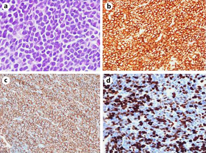
a Tissue morphology of chest wall mass demonstrating sheets of neoplastic cells. Cells were positive for CD138 (b) with aberrant CD56 expression (c) and a very high proliferative rate with Ki-67 (d).

a Tissue morphology of chest wall mass demonstrating sheets of neoplastic cells. Cells were positive for CD138 (b) with aberrant CD56 expression (c) and a very high proliferative rate with Ki-67 (d).