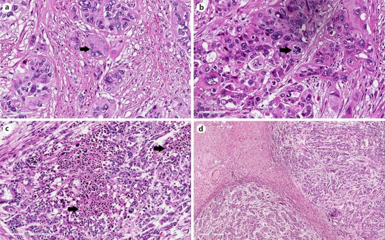
Fig. 1.

Histological findings. a Giant pleomorphic cell with micronuclei (arrow). HE. ×400. b High-power view of atypical mitosis (arrow). HE. ×400. c Low-power view of geographical necrotic areas (arrows) surrounded by viable pleomorphic cancer cells. HE. ×100. d Pushing-type neoplastic infiltrative margins. HE. ×40.
