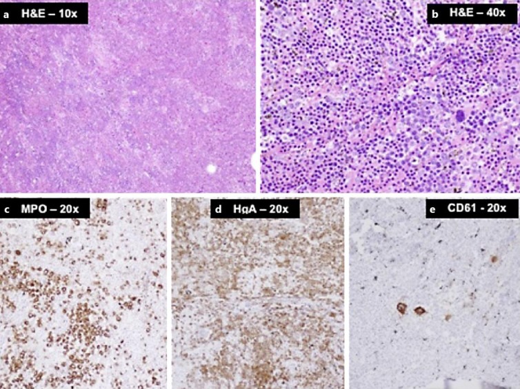
Fig. 2.

a, b Photomicrographs of tumor sections with H&E staining showing densely cellular hematopoietic marrow composed of hyperplastic erythroid elements admixed with myeloid cells and scattered megakaryocytes (large cells). c–e Immunohistochemical stains for myeloperoxidase (MPO), hemoglobin A (HgA), and CD61 highlighting the three lineages, i.e. myeloid, erythroid, and megakaryocytic cells, respectively.
