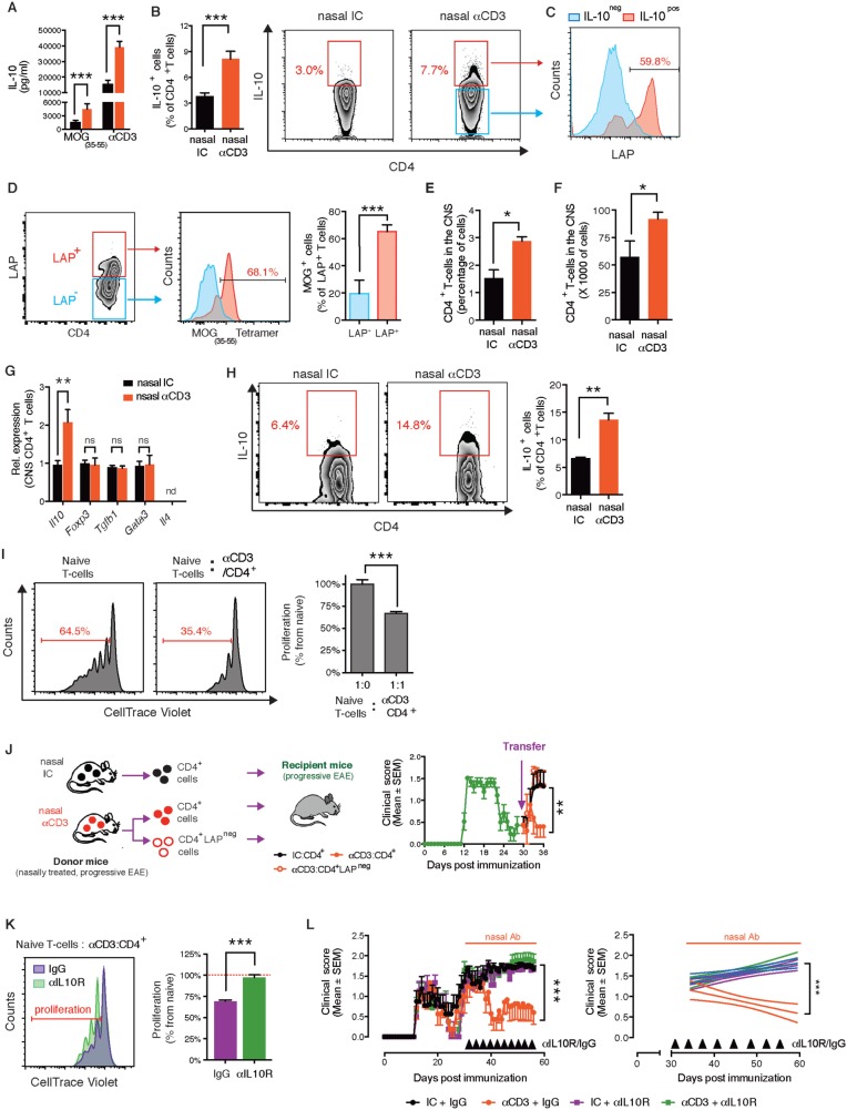
Figure 2.
Nasal anti-CD3 induces an IL-10+LAP+ T- cell that attenuates progressive EAE in an IL-10 dependent manner. ( A–I ) NOD mice treated nasally with CD3 specific or isotype control mAbs following EAE induction as in ( Fig. 1 A). IL-10 secretion by splenic T cell in response to MOG 35–55 (20 µg/ml) or anti-CD3 mAbs (0.2 µg/ml) stimulation was determined by enzyme-linked immunosorbent assay ( A ), and IL-10 and LAP expression by splenic CD4 + T cells was examined by fluorescence-activated cell sorting (FACS) ( B and C ). Data are representative of four independent experiments with n = 6 mice/group (mean and SEM). Statistical analysis by Student’s t- test. ( D ) Proportion of MOG-specific T cells within LAP + and LAP − splenic T cells in NOD EAE mice treated with nasal anti-CD3. Data are representative of two independent experiments with n = 5 mice/group (mean and SEM). Statistical analysis by Student’s t- test. Relative and absolute numbers of CNS-infiltrating CD4 + T cells was determined by FACS ( E and F , respectively). ( G ) Quantitative PCR analysis of the expression of Il10 , Foxp3, Tgfb1 , Gata3 , and Il4 mRNA of CD4 + T cells isolated from the CNS; expression is presented relative to Gapdh. ( H ) IL-10 expression by CNS-infiltrating CD4 + T cells was examined by FACS ( I and K ) FACS-sorted CD4 + T cells from nasal anti-CD3 treated NOD EAE mice (Day 60) were used in a standard suppression assay with naive CD4 + responder T cells at a ratio of 1:1 ( I ). To test the role of IL-10 in in vitro suppression, isotype control (IC) or anti-IL-10 R (50 µg/ml) blocking antibodies were added to co-cultures ( K ). Representative data of two independent experiments with n = 4 mice/group, statistical analysis by Student’s t- test. ( J ) Clinical scores of EAE in NOD mice following adoptive transfer (4 × 10 6 cells/mouse) of splenic CD4 + , or CD4 + LAP neg T cells sorted from chronic NOD EAE mice (Day 60) that were treated with anti-CD3 as in Fig. 1 A. Representative data of two independent experiments with n = 5 mice/group ( L ) Clinical scores of EAE in NOD mice. At the onset of the chronic phase of EAE mice were treated daily with nasal CD3 specific or isotype control mAbs, and were intraperitoneally injected every fourth day (black arrows) with anti-IL-10 receptor blocking mAbs (αIL-10 R) or appropriate control (IgG) (0.5 mg/mouse). Representative data of two independent experiments with n = 8 mice/group. * P < 0.05, ** P < 0.01, *** P < 0.001, n.s. = not significant; n.d. = not detected.

