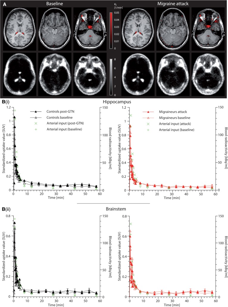
Figure 2.
The blood–brain barrier remains intact during migraine attacks . ( A ) The K i map in the upper row shows the binding pattern of 11 C-DHE overlaid on a structural MRI. The average dynamic image is depicted in the bottom row . Except for the structures outside the blood–brain barrier, i.e. choroid plexus, pituitary fossa, venous sinus, and facial tissue, there is no intracranial binding of 11 C-DHE ( K i = 0/min). There is only very low average radioactivity in the brain parenchyma. Due to the abundance of DHE receptors in the brain tissue, this indicates that 11 C-DHE is not able to cross the blood–brain barrier at baseline state ( left ). Notably, there is no change of this pattern during acute migraine attacks ( right ) suggesting that the blood–brain barrier remains intact during migraine headache. This was identical for all six migraineurs and control subjects at baseline and in post-GTN state. For illustration purposes, the K i map of one subject (Patient M6) was plotted at a threshold of K i > 0.001/min and was overlaid onto the subject’s coregistered MRI. The average dynamic PET was taken from the same subject. ( B ) The maximum penetration of the blood–brain barrier was further approximated for migraineurs ( n = 6 ) and controls ( n = 6 ) by assessing the time-activity curves (depicting mean for all time points and mean ± SEM between 10 and 60 min for the purpose of clarity) of standardized uptake values (SUV) as exemplarily shown for hippocampus [ B(i) ] and brainstem [ B(ii) ]. In all areas, the SUV followed the arterial input radioactivity (green crosses). This rapid washout of tracer supports the lack of parenchymal binding of 11 C-DHE seen in the K i maps in A . In all parenchymal areas, there was no difference between the area under the curve of the post-GTN (i.e. migraine state in migraineurs) and the respective baseline state (Wilcoxon signed-rank test: P > 0.05).

